ECV intro (張學譽 102年台科大電機所碩士論文)
心血管磁共振造影(Cardiovascular magnetic resonance, CMR)利用顯影劑延遲增強(Late Gadolinium Enhancement, LGE)影像可以顯示出心肌梗塞[1],或是利用顯影劑在細胞外的分佈顯示出非缺血性心肌纖維化疾病[2, 3],LGE影像可以容易辨別這些病灶在心肌的區域[4]。雖然LGE影像上能夠用來診斷上述心肌病灶,但卻無法用在瀰漫型心肌纖維化疾病的判讀,因為LGE影像對比是依賴正常心肌組織與心肌纖維化組織之間的信號強度差異,而瀰漫型心肌纖維化是均勻分佈在心肌組織中,所以在LGE影像上信號強度差異並不明顯[5]。瀰漫型心肌纖維化疾病主要是因為心肌細胞間的膠原蛋白含量和細胞外其它成分增多,導致心肌胞外容積(ECV)增加[6],瀰漫型心肌纖維化疾病跟高血壓性心臟病、主動脈瓣狹窄和心肌炎有關[7-9],導致呼吸困難,心臟衰竭,心律失常[10-12]。
心肌細胞外容積比(ECV)映像這項技術是在計算單位體積中心肌胞外體積所佔有的比例,正常ECV值約為25.4 ± 2.5% [13],ECV值量化計算,將會有助於心肌纖維化瀰漫型疾病的偵測,也可以用來偵測心肌梗塞和非缺血性心肌纖維化疾病[14]。利用ECV量化的範圍也可以用來評估心肌受損的情況,如果心肌纖維化程度或心肌梗塞程度上升,則ECV的值也會上升,對於用來偵測心肌疾病病灶和檢視心肌受損程度來看,ECV映像會是ㄧ個在心臟醫學影像上很有效的工具。
心肌胞外容積比(ECV)實驗假設
ECV映像的實驗是透過注射磁共振顯影劑,當顯影劑濃度達到動態平衡時計算求得細胞體積外顯影劑的分佈比例,磁共振造影中,最普遍使用的顯影劑是含釓 (Gadolinium) 顯影劑,因釓為有強力順磁性的特性,所以主要用來當核磁共振的顯影劑,其造成細胞組織的T1時間縮短,使得T1權重之磁共振影像對比增強。
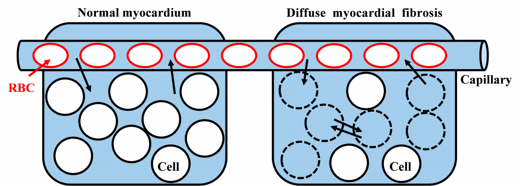
圖1-1所示,為ECV實驗假設示意圖,圖中左半邊為正常的心肌細胞組織,右半邊為有瀰漫型心肌纖維化病變的心肌細胞組織,紅色圓圈為紅血球,紅血球所在的地方為血管,黑色圓圈為心肌細胞,黑色虛線圓圈為壞死的心肌細胞。實驗假設是當注射完顯影劑經過ㄧ段時間後,血管內與細胞組織外的顯影劑濃度會達到ㄧ個動態平衡,圖中黑色箭頭讓我們知道顯影劑流動的方向,而看右半邊的心肌細胞組織,當心肌細胞壞死後顯影劑就可以流入心肌細胞內使得顯影劑所佔的區域增加。所以血管內和細胞組織外淡藍色區域範圍的大小,即為心肌胞外容積比(ECV)。
可參考下列影片來詳細了解。
參考資料
1 Kim, R.J., Fieno, D.S., Parrish, T.B., Harris, K., Chen, E.L., Simonetti, O., Bundy, J., Finn, J.P., Klocke, F.J., and Judd, R.M.: ‘Relationship of MRI Delayed Contrast Enhancement to Irreversible Injury, Infarct Age, and Contractile Function’, Circulation, 1999, 100, (19), pp. 1992-2002
2 Arheden, H., Saeed, M., Higgins, C.B., Gao, D.W., Bremerich, J., Wyttenbach, R., Dae, M.W., and Wendland, M.F.: ‘Measurement of the distribution volume of gadopentetate dimeglumine at echo-planar MR imaging to quantify myocardial infarction: comparison with 99mTc-DTPA autoradiography in rats’, Radiology, 1999, 211, (3), pp. 698-708
3 Mahrholdt, H., Wagner, A., Judd, R.M., Sechtem, U., and Kim, R.J.: ‘Delayed enhancement cardiovascular magnetic resonance assessment of non-ischaemic cardiomyopathies’, European heart journal, 2005, 26, (15), pp. 1461-1474
4 Thomson, L.E., Kim, R.J., and Judd, R.M.: ‘Magnetic resonance imaging for the assessment of myocardial viability’, Journal of magnetic resonance imaging : JMRI, 2004, 19, (6), pp. 771-788
5 Karamitsos, T.D., and Neubauer, S.: ‘Detecting diffuse myocardial fibrosis with CMR: the future has only just begun’, JACC. Cardiovascular imaging, 2013, 6, (6), pp. 684-686
6 Gazoti Debessa, C.R., Mesiano Maifrino, L.B., and Rodrigues de Souza, R.: ‘Age related changes of the collagen network of the human heart’, Mechanisms of Ageing and Development, 2001, 122, (10), pp. 1049-1058
7 Villari, M.D.P.B., Vassalli, M.D.G., Schneider, M.D.J., Chiariello, M.D.M., and Hess, M.D.O.M.: ‘Age Dependency of Left Ventricular Diastolic Function in Pressure Overload Hypertrophy’, Journal of the American College of Cardiology, 1997, 29, (1), pp. 181-186
8 Schwartzkopff, B., Frenzel, H., Dieckerhoff, J., Betz, P., Flasshove, M., Schulte, H.D., Mundhenke, M., Motz, W., and Strauer, B.E.: ‘Morphometric investigation of human myocardium in arterial hypertension and valvular aortic stenosis’, European heart journal, 1992, 13 Suppl D, pp. 17-23
9 St John Sutton, M.G., Lie, J.T., Anderson, K.R., O’Brien, P.C., and Frye, R.L.: ‘Histopathological specificity of hypertrophic obstructive cardiomyopathy. Myocardial fibre disarray and myocardial fibrosis’, British Heart Journal, 1980, 44, (4), pp. 433-443
10 Hein, S., Arnon, E., Kostin, S., Schonburg, M., Elsasser, A., Polyakova, V., Bauer, E.P., Klovekorn, W.P., and Schaper, J.: ‘Progression from compensated hypertrophy to failure in the pressure-overloaded human heart: structural deterioration and compensatory mechanisms’, Circulation, 2003, 107, (7), pp. 984-991
11 Heling, A., Zimmermann, R., Kostin, S., Maeno, Y., Hein, S., Devaux, B., Bauer, E., Klovekorn, W.P., Schlepper, M., Schaper, W., and Schaper, J.: ‘Increased expression of cytoskeletal, linkage, and extracellular proteins in failing human myocardium’, Circulation research, 2000, 86, (8), pp. 846-853
12 Villari, B., Campbell, S.E., Hess, O.M., Mall, G., Vassalli, G., Weber, K.T., and Krayenbuehl, H.P.: ‘Influence of collagen network on left ventricular systolic and diastolic function in aortic valve disease’, J Am Coll Cardiol, 1993, 22, (5), pp. 1477-1484
13 Kellman, P., Wilson, J.R., Xue, H., Ugander, M., and Arai, A.E.: ‘Extracellular volume fraction mapping in the myocardium, part 1: evaluation of an automated method’, Journal of cardiovascular magnetic resonance : official journal of the Society for Cardiovascular Magnetic Resonance, 2012, 14, pp. 63
14 Ugander, M., Oki, A.J., Hsu, L.Y., Kellman, P., Greiser, A., Aletras, A.H., Sibley, C.T., Chen, M.Y., Bandettini, W.P., and Arai, A.E.: ‘Extracellular volume imaging by magnetic resonance imaging provides insights into overt and sub-clinical myocardial pathology’, European heart journal, 2012, 33, (10), pp. 1268-1278


Recent Comments